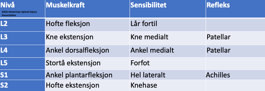
L2 (key examinations highlighted):

Hip flexion (m. iliopsoas), Sensation front of thigh.

L3 (key examinations highlighted):

Hip flexion (m. iliopsoas), L3 nerve stretch test, Sensation medial knee joint, Patellar reflex.

  • Hip adduction (m. adductor brevis, longus and magnus)

L4 (key examinations highlighted):

Dorsiflexion ankle (m. tibialis anterior), Lasegue, Sensation medial malleolus, Patellar reflex.

  • Extension big toe (m. extensor hallucis longus)

L5 (key examinations highlighted):

Extension big toe (m. extensor hallucis longus), Lasegue, Sensation between 1-2 toe.

  • Eversion forefoot (m. peroneus = fibularis)
  • Abduction hip (m. gluteus medius)

S1 (key examinations highlighted):

Plantar reflex foot (m. gastrocnemius), Lasegue, Sensation lateral edge of foot, Achilles reflex.

  • Eversion forefoot (m. peroneus = fibularis)
  • Knee flexion (hamstrings = m. semitendinosus + m. semimembranosus + m. biceps femoris)

S2 (key examinations highlighted):

Hip extension (m. gluteus maximus), Lasegue, Sensation heel.

  • Knee flexion (hamstrings)
  • Plantar flexion foot (m. gastrocnemius)

C1-S5
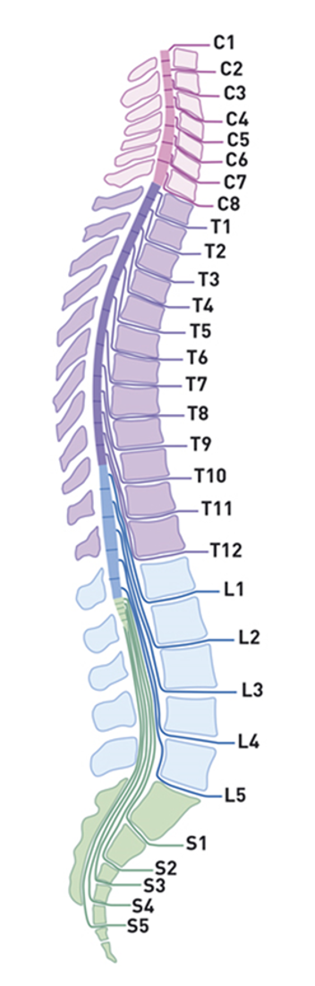
C1-S5

Tawa N, Rhoda A, Diener I. Accuracy of clinical neurological examination in diagnosing lumbo-sacral radiculopathy: a systematic literature review. BMC Musculoskelet Disord. 2017 Feb 23;18(1):93. doi: 10.1186/s12891-016-1383-2. PMID: 28231784; PMCID: PMC5324296.

Location of lumbar disc herniation in the spinal canal
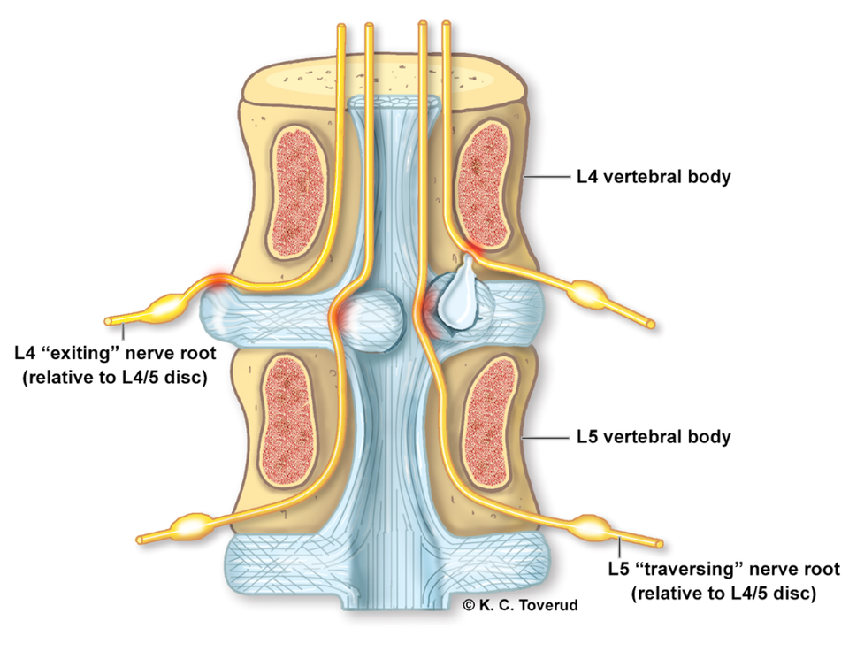
Location of lumbar disc herniation in the spinal canal
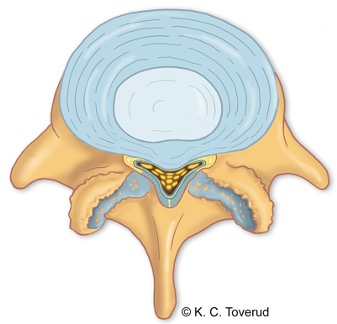
  • A paracentral (posterolateral) herniation (B) in the spinal canal at level L4/5 affects the descending (traversing) L5 nerve root, but often also the exiting L4 nerve root
  • A lateral (foraminal) herniation (A) in the spinal canal at level L4/5 usually affects only the exiting L4 nerve root
  • This depends on the anatomy of the herniation
  • The same applies at level L5/S1
  • 95% of all herniations occur at levels L4-L5 and L5-S1
  • Most flexion, extension and lateral bending in the lumbar spine takes place at these two levels

Posterolateral (paracentral) herniation is most common
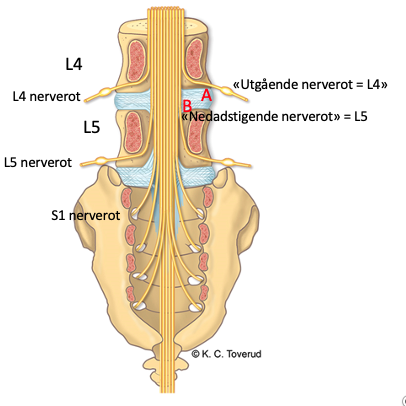
Posterolateral (paracentral) herniation is most common
  • The L4 nerve root exits through the L4-L5 neural foramen
  • The L5 nerve root exits through the L5-S1 neural foramen
  • The S1 nerve root exits through the first neural foramen of the sacrum
  • Only 10% of cases involve a lateral (foraminal) herniation (A) that presses on the "exiting nerve root"
  • An L4-L5 posterolateral (paracentral) herniation (B) will affect the L5 nerve root as the "descending nerve root", and in the same way an L5-S1 posterolateral (paracentral) herniation will affect the S1 nerve root

Tor Ingebrigtsen Behandlingsrater for ryggkirurgi i Norge og Helse Nord 2014–18

Tidsskr Nor Legeforen 2020 doi: 10.4045/tidsskr.20.0313

Lateral spinal stenosis (lateral recess stenosis)
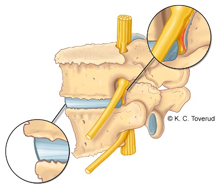
Lateral spinal stenosis (lateral recess stenosis)
  • Caused by facet joint osteoarthritis with osteophytes at level L4/5 or by a laterally located disc herniation
  • Typical symptoms are unilateral leg pain in the L4 or L5 dermatome respectively
  • Symptoms are reproduced by
    • Active lateral bending
    • Extension and rotation

Central spinal stenosis

Central spinal stenosis
  • Age group > 65 years
  • Most common at disc level L4-5, L5 nerve root
  • Narrowing of the spinal canal, A-P diameter > 10 mm, due to osteoarthritis, ligament thickening or disc bulging
  • Moderate symptoms at first, later spinal claudication and short walking distance with calf pain
  • Symptoms are relieved by bending forwards or sitting leaning forwards
  • Few clinical neurological findings on examination

Neurogenic versus vascular claudication

FunctionNeurogenic claudicationVascular claudication
WalkingCauses painCauses pain
StandingCauses painSymptom relief
SittingSymptom reliefSymptom relief
StairsEasier going upEasier going down
CyclingSymptom reliefCauses pain
Ankle pulseNormalWeakened

Foot drop, several nerve roots affected
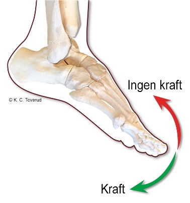
Foot drop, several nerve roots affected

In a study by Liu (2013) of 62 patients with foot drop due to disc herniation, 25/62 (40.3%) had involvement of several nerve roots, in various combinations of (L4) + L5 + (S1), with the L5 root most often involved

Liu K, Zhu W, Shi J, Jia L, Shi G, et al. (2013) Foot Drop Caused by Lumbar Degenerative Disease: Clinical Features, Prognostic Factors of Surgical Outcome and Clinical Stage. PLoS ONE 8(11): e80375. doi:10.1371/journal.pone.0080375

Differential diagnosis of foot drop

  • Foot drop may be caused by radiculopathy (disc herniation compressing the L4 and L5 nerve roots) or neuropathy (pressure on the N. fibularis (preoneus) communis)
  • M. tibialis anterior, M. extensor digitorum longus, M. hallucis longus, Mm. fibularis (peroneus) longus and brevis dorsiflex and evert the ankle. The nerve supply is via the N. fibularis (peroneus) communis
  • In foot drop caused by neuropathy of the N. fibularis (peroneus) communis, inversion strength is normal
  • Foot drop caused by L5 radiculopathy shows reduced strength in dorsiflexion, eversion and inversion. M. tibialis posterior inverts the ankle and is innervated by the N. tibialis. Together with foot drop, reduced hip abduction strength can often be demonstrated because of weakness of M. gluteus medius (L5 innervated). Radiculopathy also causes numbness and radiating pain in the L5 dermatome.
Back to the back